Mammograms
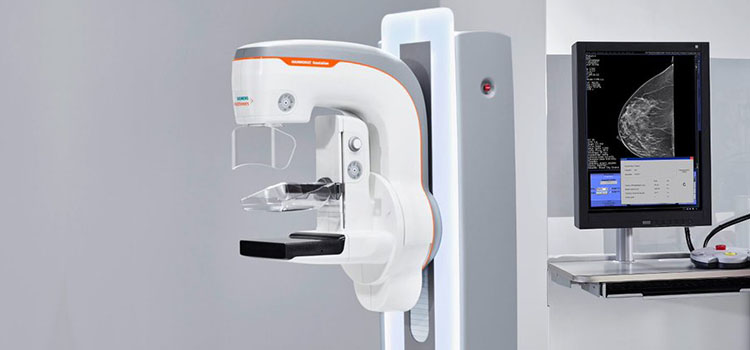
Mammography uses low doses of X-rays to examine the breast accurately. It is the most effective tool for the earliest detection of breast cancer because mammography can reveal masses in the breast up to two years before a patient or physician can feel them.
Before scheduling a mammogram, discuss any problems or concerns you have with your breasts with your doctor. Inform your doctor of any hormone use, prior surgeries, and any family or personal history of breast cancer.
If you have sensitive breasts, the best time to have a mammogram performed is one week following your period. Try not to have your mammogram during the week before your period if your breasts are typically tender during this time. Please also inform your mammography technologist if there is any possibility that you are pregnant.
What is Mammography?
mammogram is a low dose x-ray image of the breast. It is the most common and cost-effective screening exam for breast cancer and for diagnosing a variety of breast issues.
Until recently, all mammography used 2 dimensional (2D) images, which captured all of the breast tissue in 4-6 images.
What is 3D mammography, and why do we recommend 3D Mammography for our patients?
Types of Mammograms
Screening Mammogram
A routine annual exam recommended for women who do not have breast symptoms. Screening mammograms aim to detect unsuspected breast cancers at an early stage when treatment is most effective.
Diagnostic Mammogram
We perform diagnostic mammograms when a woman or health care professional discovers a lump, tenderness, nipple discharge or skin changes - or after a patient has had suspicious results on a screening mammogram.
We also perform Diagnostic mammograms when your practitioner recommends a short term follow-up from a prior diagnostic mammogram, or if a patient had breast cancer.
We interpret diagnostic mammogram in a few ways:
- The area on a screening mammogram is normal, and no further evaluation is needed.
- The area is not cancerous, but the radiologist recommends following the area with a four to six-month diagnostic follow up.
- The area of concern that is suspicious for cancer and suggests a biopsy for confirmation.
When will I get my results?
The referring physician will receive a copy of the finalized report within 24 hours.
Breast Ultrasound
A Breast Ultrasound is an imaging modality commonly used to screen or evaluate tumours in the breast. Ultrasounds use high-frequency sound waves to produce images of the inside of the breast. Because ultrasound does not use radiation, it is considered safe for pregnant or breastfeeding patients.
When is a breast ultrasound performed?
Referring physicians may recommend an ultrasound for patients that are under the age of 30, are pregnant, are breastfeeding or have silicone breast implants. Physicians may also recommend a patient have an ultrasound and mammogram if they feel a palpable lump on a breast exam.